Prepare for a voice-first exam
Use a lapel microphone for a typical 10 to 20 minute chairside exam. Confirm that the microphone is connected and positioned before the clinical conversation begins.
Have relevant evidence ready, such as:
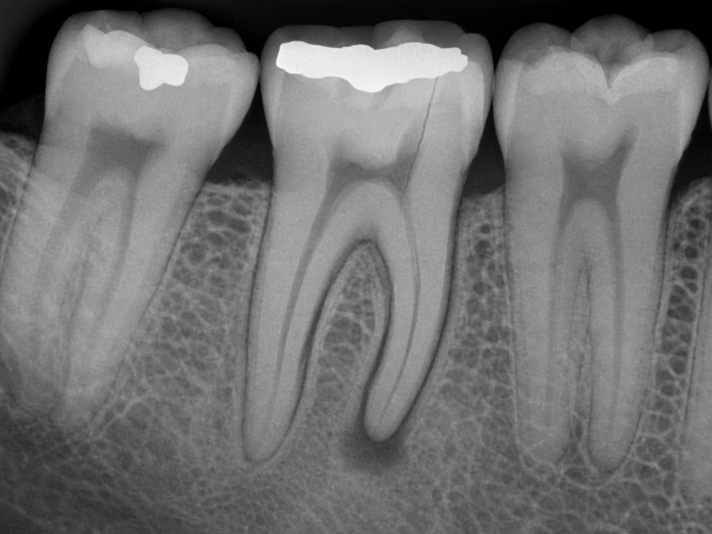
- Radiographs.
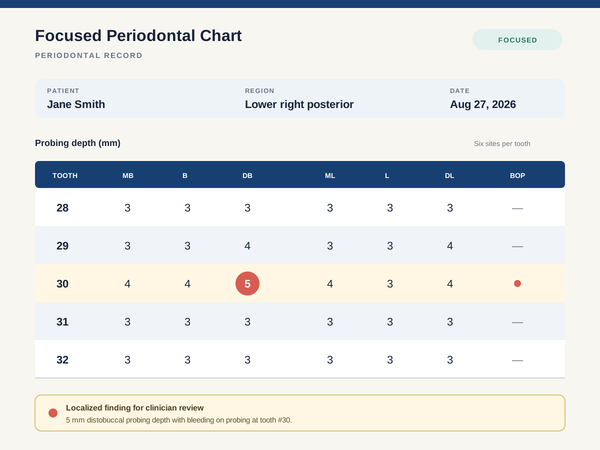
- Periodontal charts.
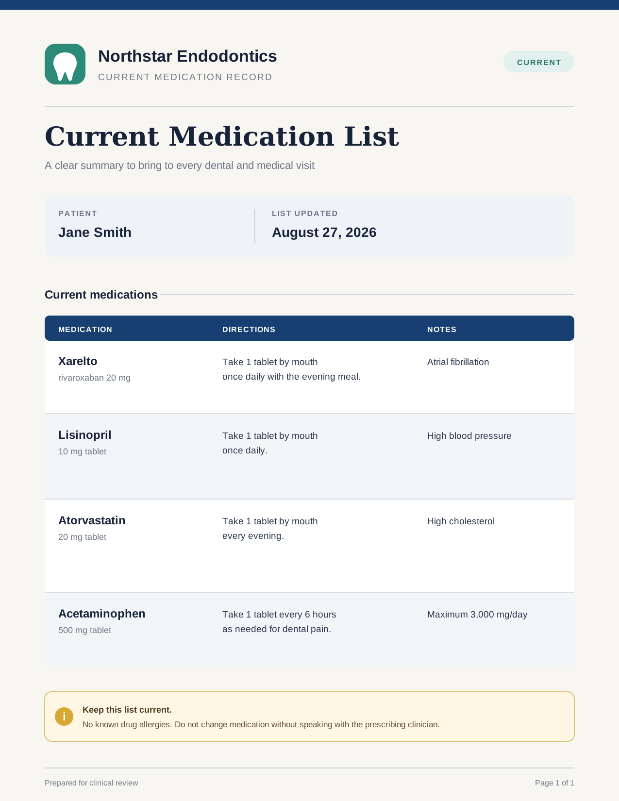
- Health-history forms.
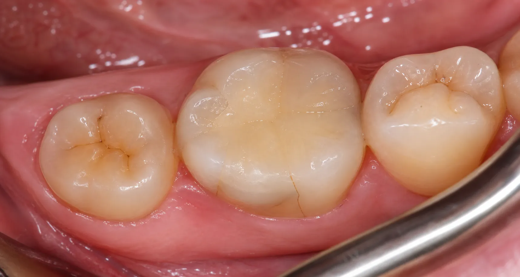
- Clinical photographs.
- Existing patient records.
Primary source
Chairside exam recording
 OcclusalCrack detail
 BuccalPerio site
Select the signing clinician
Choose the clinician responsible for the encounter before recording. This determines who signs the resulting documents and which applicable Voice settings are used.
In a multi-clinician organization, recording cannot begin until the signing clinician is selected.
Record the real exam
Start the recording and conduct the exam normally. Speak to the patient and team in the way you already work, while making the clinically important information audible.
Narrate:
- The patient's reason for the visit and relevant history.
- Clinical and radiographic findings.
- Your assessment.
- The treatment discussion.
- Recommendations and next steps.
You do not need to dictate a SOAP template or announce punctuation.
Add supporting evidence
Attach relevant documents before generation. ToothNote.ai can use the recording alongside radiographs, periodontal charts, health-history forms, photographs, and patient records.
The completed encounter keeps the original recording and transcript available for review. The sources remain connected to the case rather than becoming separate document fragments.
Review extracted context
Before relying on generated documents, review the supporting context ToothNote.ai found:
- Radiographic findings presented as documentation support.
- Periodontal-chart findings that require clinical confirmation.
- Medical and dental-history context.
- Information from the recording and transcript.
Supporting evidence
Proposed context for clinician confirmation
Medical considerations
Rivaroxaban 20 mg daily for atrial fibrillation; lisinopril and atorvastatin listed; no known drug allergies.
Periodontal findings
Localized 5 mm distobuccal probing depth with bleeding at #30; remaining focused sites measure 3–4 mm.
Radiographic findings
Large restoration at #30, widened PDL space, and small periapical radiolucency at the mesial root.
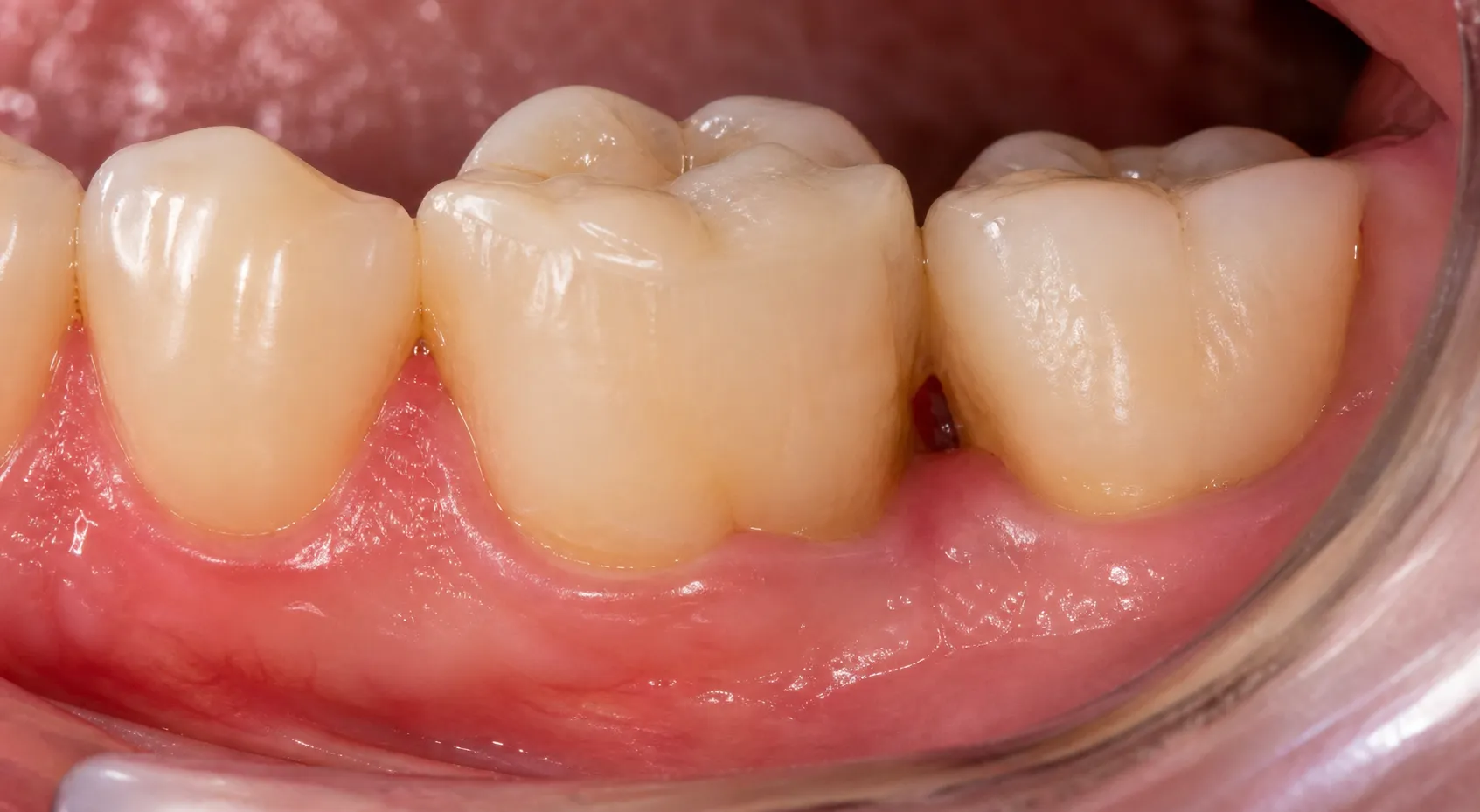
Photographic findings
Distal marginal-ridge crack adjacent to the MOD composite; mild localized gingival inflammation without visible swelling.
Review the three core outputs
One complete exam produces three core documents for three different audiences.
SOAP Clinical Note
Focused endodontic evaluation · Tooth #30
Subjective
58-year-old patient referred for intermittent lower-right biting pain for three weeks. Cold sensitivity lingers after the stimulus is removed. No spontaneous swelling or drainage reported.
Objective
#30 has a large MOD composite and visible distal marginal-ridge crack. Cold response is exaggerated and lingers 18 seconds; percussion positive; palpation negative. Localized 5 mm distobuccal probing with bleeding. Radiograph shows widened PDL space and a small mesial-root periapical radiolucency.
Assessment
Symptomatic irreversible pulpitis with symptomatic apical periodontitis, tooth #30. Restorability to be confirmed after removal of the existing restoration and evaluation of the crack.
Plan
Discussed nonsurgical root canal treatment, prognosis, alternatives, and the need for a definitive cuspal-coverage restoration. Medication history, including rivaroxaban, reviewed; no medication changes advised by the dental team.
Reviewed clinical note
Consultation Report
Re: Endodontic evaluation of tooth #30
Dear Dr. Rivera,
Thank you for referring our shared patient for evaluation of tooth #30. The patient described three weeks of pain when biting and lingering cold sensitivity.
Findings
Clinical testing was consistent with symptomatic irreversible pulpitis and symptomatic apical periodontitis. A distal marginal-ridge crack is visible adjacent to the existing MOD composite. The periapical image shows widened PDL space and a small radiolucency at the mesial root.
Recommendation
We recommended nonsurgical root canal treatment on #30, with restorability confirmed after the existing restoration is removed. If the crack is confined coronally, the patient should return to your office promptly for cuspal-coverage restoration.
Sincerely,
Alex Morgan, DDS · Northstar Endodontics
Reviewed consultation report
Your Visit Today
A plain-language summary of your lower-right molar exam
What we found
The nerve inside your lower-right first molar (#30) is inflamed and is not expected to heal on its own. We also saw a small crack next to the large filling in that tooth.
Recommended next step
Root canal treatment can remove the inflamed tissue and help you keep the tooth. During treatment, we will evaluate how far the crack extends. Afterward, your general dentist will likely protect the tooth with a crown or other cuspal-coverage restoration.
Until your appointment
Avoid chewing hard foods on the lower right. Continue medications only as prescribed. Call us sooner if you develop facial swelling, fever, drainage, or rapidly increasing pain.
Questions?
We are happy to review the findings, options, expected sequence, and costs with you before treatment.
Patient-ready visit summary
Edit, approve, and deliver
Review each document for its own audience. The same encounter supports all three, but the actions and recipient are different.
Clinical Note
- Edit individual sections or regenerate the note.
- Save the approved note as a writing sample.
- Copy plain text into the PMS.
- Download HTML, PDF, DOCX, or JPG.
- Send it to your inbox or assigned staff.
Referral Letter
- Change the signer, referring doctor, or date.
- Regenerate or save the approved letter as a writing sample.
- Include selected radiographic evidence.
- Copy plain text or download PDF, HTML, DOCX, or JPG.
- Email the referring doctor when an address is on file.
Patient Take-Home
- Edit or regenerate the plain-language recap.
- Copy the approved content.
- Download the branded PDF, DOCX, or HTML document.
- Email the branded PDF when the patient has an address on file.
PDF, HTML, DOCX, JPG, copy, download, and email are Delivery choices. They are not Format settings.
Create optional follow-on documents
When the case requires them, the reviewed encounter can also begin:
These are optional downstream workflows. They are not automatic core outputs, and they do not replace the letter back to the doctor who originally referred the patient.
Complete the workflow
Approving the three core outputs may not automatically close the entire case workflow. Complete any needed specialist referral or medical-clearance work.
Skip optional branches that do not apply, or choose Mark complete anyway when the case is finished without them.